Office-Based Procedures
Lake Medical Imaging provides office-based procedures that often replace traditional surgery.
Venous Ablation: Varicose Veins
Venous ablation is performed in our office using imaging guidance. After applying local anesthetic to the vein, one of our interventional radiologists inserts a thin catheter, about the diameter of a strand of spaghetti, into the vein and guides it up the greater saphenous vein in the thigh. Then laser energy is applied to the inside of the vein. This heats the vein and seals it. By closing the greater saphenous vein, the twisted and varicosed branch veins, which are close to the skin, shrink and leg appearance improves. Once the diseased vein is closed, other healthy veins take over to carry blood from the leg, thereby re-establishing normal flow.
Benefits of Venous Ablation Treatment
You may return to normal activity the next day with little or no pain (there may be minor soreness or bruising, which can be treated with over-the-counter pain relievers). Because the procedure does not require a surgical incision – simply a nick in the skin about the size of a pencil tip – there are no scars or stitches. Traditionally, surgical ligation or vein stripping was the treatment for varicose veins, but these procedures can be quite painful and often have a long recovery time. The success rate for venous ablation is substantially higher than that for surgery (meaning the recurrence rate is low); in fact, the success rate for venous ablation ranges from 93% to 95%.
Sclerotherapy: Spider Veins
Sclerotherapy is the most common treatment for spider veins. The procedure takes about one hour, during which time a solution called a “sclerosing agent” is injected into the veins. This causes the inter-lining of the vein to adhere, which results in scar tissue that acts like ‘glue’ to seal off the unwanted veins. There are a few side effects, such as bruising around the injection site, or temporary discoloration of a light brown color along the vein. However, you may resume normal activity immediately.
How many treatments will it take?
The number of treatments depends on the individual and extent of the spider veins; one or possibly more treatments. Typically, the average number of treatments required are three.

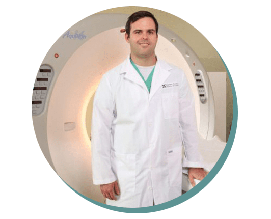
Kyle Hopkins, PA-C

J. Laird McMullen, III, M.D.
Image-Guided Needle Biopsy
In many cases, tissue samples can be obtained without open surgery by utilizing interventional radiology techniques. Nearly all biopsies performed by interventional radiologists are image-guided needle biopsies. This means that a needle is placed into an area of abnormality directly through the skin. The needle placement is confirmed with the use of a variety of equipment used by the radiologists including ultrasound (used most often), CT or x-ray fluoroscopy.
Needle biopsies are almost always performed as an outpatient procedure with a short (one hour) observation period thereafter.
Paracentesis and Thoracentesis
Patients with a variety of illnesses may develop an area of excessive fluid in a body cavity, such as the abdomen or chest. The fluid can be drained by inserting a thin tube (catheter) through a small nick in the skin. In an effort to determine the cause of the fluid build-up or to relieve the symptoms of the excess fluid, interventional radiologists insert a small tube (less than 1/12 of an inch in diameter) through the skin, after numbing the skin with a local anesthetic. The fluid may be sent to the laboratory for analysis.
Planning for your Paracentesis and Thoracentesis procedure
An ultrasound will be used to help decide where to insert the needle. Your skin will be cleaned and the area around the procedure site covered with a clean sheet. Local anesthesia makes you more comfortable during your procedure and is used to numb the insertion area and dull your pain. However, you may still experience pressure or pushing during the procedure.
A needle will be inserted into your abdominal or chest cavity. A catheter is attached to the needle and the needle is removed. The catheter tubing will be attached to a container with a suction device (a gentle vacuum) to help drain the fluid. Removing large amounts of fluid may take a couple hours. After the procedure is completed and the catheter removed, the site will be covered with a bandage. Your referring physician may request that the ascites or pleural fluids be sent to a lab for tests.
Once the procedure is completed and the catheter removed, the site will be covered with a bandage. Your referring physician may request that the ascites or pleural fluids be sent to a lab for tests.
A catheter approximately the size of a pen tip is guided under fluoroscopy into the desired vessels in your liver. Ionizing radiation via millions of tiny glass beads is then delivered into the tumor.
For more information, visit: Radiologyinfo.org Thoracentesis

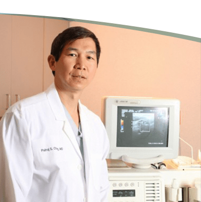
Pairoj Sae Chang, M.D. Ph.D.
Vertebral Augmentation
Vertebral Augmentation (VA) is a procedure performed by an interventional radiologist in order to reduce the discomfort associated with vertebral body fractures in the spine. These fractures are typically secondary to osteoporosis; however, sometimes these fractures can be caused by trauma or tumors. Vertebral body fracture is often associated with vertebral body height loss. Studies have shown that if the vertebral body height loss can be improved, then there is less associated morbidity, such as pain and decreased mobility.
At the present time, Medicare and other third-party insurers authorize vertebral augmentation for pain management. Both vertebroplasty and kyphoplasty are outpatient procedures that also can be performed on an inpatient basis. Vertebroplasty is a delivery of an orthopaedic “cement” (polymethylmethacrylate) mixed with barium. Under fluoroscopy or CT guidance, the orthopaedic cement is administered into the fractured vertebral body through placement of one to two needles. There is often no height restoration of the fractured vertebral body.
Kyphoplasty is a delivery of PMMA mixed with barium into the vertebral body after balloon dilation of the vertebral body. There are other forms of kyphoplasty that can improve the height of a compressed vertebral body. VA can be safely performed on up to three fractured vertebral bodies at any given time.
Planning for your procedure
Vertebroplasty:
The interventional radiologist will review your imaging studies and he or she may require additional imaging studies and/or a consultation. You may be asked to stop certain anticoagulants several days prior to the procedure, and may be directed to consult the physician who prescribed them. You may continue to take your other medications, as usual. Eight hours prior to the procedure, stop eating solid foods. You may continue drinking liquids up to four hours prior to the procedure and taking your oral medications with few sips of water up to an hour prior to the procedure.
On the day of the procedure, wear loose comfortable clothing. Arrange for a designated driver to drive you home post-procedure.
An IV is started and an antibiotic administered pre-procedure. You are escorted to a fluoroscopy suite where the procedure is performed. You lie on your stomach and conscious sedation is administered intravenously. After sterile prep of your back, a local anesthetic is administered. Under fluoroscopy, two needles are placed – one through each pedicle of the fractured vertebral body while the orthopaedic cement is introduced. The needles are then removed and adhesive bandages are placed over the puncture sites. For one level, the procedure time is approximately 30 minutes. For two or three levels, the procedure time is approximately 60 minutes.
Depending on how quickly you recover from the sedation post-procedure, recovery is one to two hours. You may experience complete pain relief; however, some pain may return after 12 to 24 hours. The immediate pain relief may be due to the long-acting local anesthetic. A 50% pain relief post-procedure is considered a success. Arrange for a designated driver to drive you home. During your first night home, we prefer that you are not alone and have an adult with you. You are required to schedule a consultation with us two to three weeks post procedure, so that we may assess your progress.
A catheter approximately the size of a pen tip is guided under fluoroscopy into the desired vessels in your liver. Ionizing radiation via millions of tiny glass beads is then delivered into the tumor.
Kyphoplasty:
The interventional radiologist will review your imaging studies and may require additional imaging studies and/or a consultation. You may be asked to stop certain anticoagulants several days prior to the procedure. Before you stop taking your anticoagulants temporarily, you may be required to consult with the physician who prescribed them. You may continue to take your other medications as usual. Eight hours prior to the procedure, stop eating solid foods. You may continue drinking liquids up to four hours prior to the procedure and taking your oral medications with few sips of water up to an hour prior to the procedure.
On the day of the procedure, wear loose comfortable clothing. Arrange for a designated driver to drive you home post-procedure.
An IV is started and an antibiotic administered pre-procedure. You are escorted to a fluoroscopy suite where the procedure is performed. You lie on your stomach and conscious sedation is administered intravenously. After sterile prep of your back, a local anesthetic is administered. Under fluoroscopy, two needles are placed – one through each pedicle of the fractured vertebral body – and the balloon device deployed followed by the “cement”. The needles are then removed and adhesive bandages are placed over the puncture sites. For one level, the procedure time is approximately 45 minutes. For two or three levels, the procedure time is approximately 60 to 90 minutes.
Depending on how quickly you recover from the sedation post-procedure, recovery is one to two hours. You may experience complete pain relief; however, some pain may return after 12 to 24 hours. The immediate pain relief may be due to the long-acting local anesthetic. A 50% pain relief post-procedure is considered a success. Arrange for a designated driver to drive you home. During your first night home, we prefer that you are not alone and have an adult with you. You are required to schedule a consultation with us two to three weeks post procedure, so that we may assess your progress.
A catheter approximately the size of a pen tip is guided under fluoroscopy into the desired vessels in your liver. Ionizing radiation via millions of tiny glass beads is then delivered into the tumor.
Diagnostic Biopsies
These procedures are typically requested by referring physicians. The interventional radiologist then reviews the previously obtained imaging study and determines the need and feasibility of a minimally invasive biopsy. We perform these procedures under imaging guidance (US, CT, fluoroscopy, or MRI).
Lung Biopsy
These procedures are typically requested by referring physicians. The interventional radiologist then reviews the previously obtained imaging study and determines the need and feasibility of a minimally invasive biopsy. You will be advised to stop taking aspirin, non-steroidal anti-inflammatory drugs (NSAIDs) or blood thinners several days prior to your procedure. We perform these procedures under CT guidance.
The interventional radiologist will clean the area of skin the needle will go through, place a sterile drape around the area and numb the area. Using CT guidance, a needle will then be inserted through the skin and into the lung to the nodule and multiple samples will be taken with the samples sent to pathology for evaluation. After the procedure, you will be observed by nurses for routine monitoring. You may experience pressure during tissue sampling as well as an urge to cough. You may experience the urge to cough including coughing a small amount of blood during the procedure.
You will be observed/monitored over a timeframe of one hour and will then be permitted to go home. If you receive conscious sedation, you must arrange for a designated driver to drive you home post-procedure. Your referring physician will receive the results in a few days. Very few patients may experience an air leak due to the needle causing a hole in the lung (pneumothorax). This usually heals on its own and will not require further procedures. But if the air leak is big enough, or you experience symptoms due to the air leak, a tube may need to be inserted through the skin and chest wall to drain the air from your chest cavity.
Thyroid Nodule Fine Needle Aspiration (FNA)
These procedures are typically requested by referring physicians. The interventional radiologist then reviews the previously obtained imaging study and determines the need and feasibility of a minimally invasive biopsy. You will be advised to stop taking aspirin, non-steroidal anti-inflammatory drugs (NSAIDs) or blood thinners several days prior to your procedure. We perform these procedures under ultrasound guidance.
The interventional radiologist will clean the area of skin the needle will go through, place a sterile drape around the area and numb the area. Under ultrasound guidance, the small needle will be advanced through the skin and into the nodule. Aspiration is performed with this needle attached to a syringe with gentle aspiration technique. 3-4 separate samples are typically obtained and the sample is sent to pathology for evaluation.
You will be given an ice pack to decrease any potential swelling. Patients typically experience little to no pain but may have a small amount of bruising at the biopsy site. You can resume normal activities after thyroid aspiration.
Liver Biopsy
These procedures are typically requested by referring physicians. You will be advised to stop taking aspirin, non-steroidal anti-inflammatory drugs (NSAIDs) or blood thinners several days prior to your procedure. We perform these procedures under ultrasound or CT guidance.
These procedures are typically requested by referring physicians. You may be advised to stop blood thinners several days prior to your procedure. We perform these procedures under fluoroscopic (X-ray) or CT guidance, targeting a mass or obtaining tissue anywhere from the liver to evaluate overall liver function (random liver biopsy).
You will be observed/monitored over a timeframe of one hour and will then be permitted to go home. If you receive conscious sedation, you must arrange for a designated driver to drive you home post-procedure. Your referring physician will receive the results in a few days. You may have some pain where the needle entered your skin. You may also have pain in your shoulder. It's caused by pain travelling along a nerve that goes to the liver. This pain usually lasts less than 8-12 hours.
Kidney Biopsy
These procedures are typically requested by referring physicians. You will be advised to stop taking aspirin, non-steroidal anti-inflammatory drugs (NSAIDs) or blood thinners several days prior to your procedure. We perform these procedures under ultrasound or CT guidance.
The interventional radiologist will clean the area of skin the needle will go through, place a sterile drape around the area and numb the area. Under ultrasound or CT guidance, the small needle will be advanced through the skin and into the kidney, targeting a mass or obtaining tissue from the outer portion of the kidney (cortex) to evaluate overall kidney function (random kidney biopsy).
You will be observed/monitored over a timeframe of one hour and will then be permitted to go home. If you receive conscious sedation, you must arrange for a designated driver to drive you home post-procedure. Your referring physician will receive the results in a few days. You may have some pain in the flank region where the biopsy is performed. The pain usually lasts less than 8-12 hours.
Bone Marrow Biopsy
These procedures are typically requested by referring physicians. You may be advised to stop blood thinners several days prior to your procedure. We perform these procedures under fluoroscopic (X-ray) or CT guidance.
The interventional radiologist will clean the area of skin the needle will go through, place a sterile drape around the area and numb the area. The bone biopsy needle is advanced through the outer part of the bone (cortex) to obtain marrow. Bone marrow fluid (aspirate) and tissue sample (biopsy) are usually collected from the top ridge of the back of a hipbone (posterior iliac crest).
Depending on the pain medicine required to perform the procedure (local anesthetic or local anesthetic with IV sedation), you will be monitored for a short time period and then discharged home. Make arrangements to have a ride home and plan for light activity for the remainder of day. You may experience pain in the bony pelvic region where the biopsy was performed. The pain usually lasts less than 8-12 hours.
Soft Tissue/Lymph Node Biopsy
These procedures are typically requested by referring physicians. You may be advised to stop blood thinners several days prior to your procedure. We perform these procedures under ultrasound or CT guidance depending on the location of the lesion of concern.
Your biopsy experience will depend on the location of the lesion. You may be lying on your back or stomach and you may/may not require IV sedation in addition to local anesthetic. In all cases, the interventional radiologist will clean the area of skin the needle will go through, place a sterile drape around the area and numb the area. Under ultrasound or CT guidance, the small needle will be advanced through the skin and into the lesion of concern. Tissue sampling will be performed and the samples will be sent to pathology for evaluation.
Depending on the pain medicine required to perform the procedure (local anesthetic or local anesthetic with IV sedation), you will be monitored for a short time period and then discharged home. Make arrangements to have a ride home and plan for light activity for the remainder of day.
Planning for your Diagnostic Biopsy procedure
You may be asked to stop taking blood thinning medication.
You are escorted into an imaging suite, where local anesthetic is administered. Using imaging guidance, a needle is inserted into the lesion of concern and several tissue samples are obtained.
The specimens are submitted to a lab or pathologist for diagnosis.
In some cases, an IV sedative may be administered for additional comfort.
You will be observed/monitored over a timeframe of a few minutes to a couple of hours and will then be permitted to go home. If you receive conscious sedation, you must arrange for a designated driver to drive you home post-procedure. Your referring physician will receive the results in two to three days. If the biopsy is requested by your interventional radiologist, we will contact you directly with the results.
Gastrointestinal Intervention
Interventional Radiologists will place feeding tubes into the stomach with an approach through the skin in the upper abdomen. This is usually performed in patients with difficulty swallowing related to numerous conditions including stroke, obstructive cancer of the head and neck or chronic illnesses creating an inability to maintain adequate nutrition by mouth.
You will be advised to stop taking aspirin, non-steroidal anti-inflammatory drugs (NSAIDs) or blood thinners several days prior to your procedure and instructed to not eat nor drink anything for several hours beforehand. Leave jewelry at home and wear loose, comfortable clothing. Arrange for a designated driver to drive you home post-procedure or these arrangements will be made between our nurses and your caretaker ahead of time.
In addition to local anesthetic numbing the skin, an intravenous (IV) sedative will make you feel relaxed, drowsy and comfortable for the procedure. Oftentimes, a small catheter will be placed into your stomach through your mouth in order to expand the stomach with air to allow needle passage into the stomach through your skin in the upper abdomen.
- You’ll feel some pain around the tube entry site after a percutaneous endoscopic gastrostomy. Or you might have cramping from gas buildup in your digestive system. This pain should decrease within 24 to 48 hours. You may see some drainage around the catheter entry site for up to 48 hours. Your doctor will arrange a dietician, a specialist to explain how to use the tube and start you on required nutritional regimen.
Vascular Access Procedures:
PICC Line Placement
Many patients are given temporary PICC lines to reduce the number of needle punctures during chemotherapy or antibiotic treatment or diagnostic blood draws. A vascular access procedure is designed for patients who need intravenous (IV) access for a considerable time, longer than 7 to 10 days. A simple IV set-up is effective in the short term, but is far from ideal when, for instance, a patient needs a course of chemotherapy, several weeks of IV antibiotic treatment, or long-term IV feeding.
Planning for your PICC Line Placement procedure
You may be instructed to not to eat or drink anything several hours prior to your procedure. Leave jewelry at home and wear loose, comfortable clothing. This procedure can be performed on an in-patient or out-patient basis, but if you receive conscious sedation, you should still arrange for a designated driver to drive you home afterward.
You will be positioned on your back on an angio/fluoroscopy table. A technologist may insert an intravenous (IV) line into a vein in your hand or arm so that sedative medication can be given intravenously prior to the start of or during the procedure, as needed. Your physician will numb the area with a local anesthetic. A tiny incision is made at the site. To place the PICC line and with the aid of fluoroscopy, the physician will identify the vein using ultrasound or x-ray guidance and insert a small needle into the arm vein and advance a small guide wire into the large central vein, called the superior vena cava. The catheter is then advanced over the guide wire and moved into position. The guide wire is then removed.
It is especially important to keep the catheter site clean and dry. You may be allowed to shower by wrapping a piece of watertight plastic seal wrap over the site where the catheter was inserted. You should not allow the incision to be held under water such as by swimming or soaking in a tub.
Port Placement
Ports are often requested for chemotherapy or antibiotic infusions in order to prevent venous injury. They provide a relatively safe and easy access by the healthcare providers. After the initial healing process (when the port is not accessed), you can take showers or immerse the site in a pool of water without being concerned about infection. Ports can be kept even after the initial chemotherapy has been completed. The port needs to be flushed occasionally in order to maintain the patency of the port and its catheter. You can always request a consultation with your interventional radiologist before having a port placed.
Planning for your Port Placement procedure
You may be asked to stop taking certain blood thinners for a short time before and after the port placement, in order to prevent bleeding as a result of the procedure. Continue to take your other medications, if required. You may require a blood draw in order to assess your coagulation factors.
Leave jewelry at home on the day of your procedure. You will receive an IV access and be administered intravenous antibiotics, after which time you will be escorted into a fluoroscopy suite and sterile procedure room where you will be sedated. After sterile prep and use of local anesthetic, a 4 to 5 cm incision will be made on your upper left or right chest as well as a 1 cm incision for access into your jugular vein on the same side. The port will be placed under your skin and the catheter from the port will go into your jugular vein. Incisions will be closed and dressings placed on the incision sites. You will be able to go home one or two hours after the procedure. Arrange for a designated driver to drive you home, since you will receive conscious sedation during the procedure.
You may shower, but until the incisions are healed, no baths or immersion of the incision sites into a pool of water are allowed. You may remove the clear and gauze dressings after two days, but allow the white strips of paper tape (Steristrips) to fall off on their own. The port can be accessed immediately after placement. Do not immerse the port site in a pool of water while the port is accessed with a needle.
A catheter approximately the size of a pen tip is guided under fluoroscopy into the desired vessels in your liver. Ionizing radiation via millions of tiny glass beads is then delivered into the tumor.
For more information, visit: Society of Interventional Radiology/Patient Center


