PET/CT Scan & Nuclear Medicine
PET/CT Scan
Positron Emission Tomography, also called a PET scan, is a Nuclear Medicine exam that produces a 3D image of functional processes in the body. Positron Emission Tomography (PET) scans use radio-active tracers injected intravenously (IV) to obtain images of the human body’s function and reveal information of health and disease. As the tracers journey through the body, the scanner records signals that the tracers emit and the targeted organs collect. A computer then interprets the signals and merges them with CT images, which reveal biological maps of organ function.
This type of imaging is some of the most advanced available to diagnose cancers and other diseases. PET scans are also utilized to examine the effects of cancer therapy by characterizing biochemical changes in the cancer. To determine if they are candidates for surgery, patients who have suspected or proven brain tumors, or who have seizure disorders that are not responsive to medical therapy can be evaluated using brain scans. Patients who have memory disorders of an undetermined cause are not candidates for surgery.
Patients lie on a table that moves slowly through the large opening of the PET scanner. Scan times usually average about 30 minutes, but vary depending on the region being assessed.
Planning for your PET/CT procedure
You should:
- Wear comfortable clothes with no metal zippers or buttons
- Avoid exercise and not engage in any form of strenuous or vigorous activity for 24 hours before your exam; for example, golfing, swimming, heavy lifting and housework
Any additional preparation for this test varies depending on the purpose of the scan. Expect a call from our preregistration staff who will instruct you on how to prepare for your exam.
- Plan on being at the office for 1 ½ to 2 hours (registration, injection and scan duration are included in the time approximation)
- You will receive an intravenous (IV) injection of the radio-active pharmaceutical
- The radio-active tracer will take approximately 60 minutes to travel through your body and be absorbed by the tissue under study. During this time, you will be in a comfortable recliner and private room. You will be asked to rest quietly and avoid significant movement or talking, which may alter the localization of the administered substance
- You will be positioned on the PET scanner table and asked to lie still during your exam
- The table will move slowly through the large opening of the PET scanner
- Scanning of the brain takes less than ten minutes. Scanning of the body takes between 15 and 25 minutes
- Usually, there are no restrictions on daily routine after the test. You should drink plenty of liquids to flush the radio-active substance from your body
What will I experience during the procedure?
- When given the intravenous injection, you will feel a slight prick. However, you will not feel the substance in your body
- You will be made as comfortable as possible on the exam table before you are positioned in the PET scanner for the test
- Your technologist can see, hear and speak with you at all times during your scan
The small amount of radiotracer in your body will lose its radio-activity over time through the natural process of radio-active decay. Following the test, it may also pass out of your body through your urine or stool during the first few hours or days. You should not feel any side effects and be able to return to your normal daily activities directly after the scan. We recommend that you hydrate thoroughly for 48 hours after the scan to help void the medications.
Keep your distance from children and pregnant women for approximately six hours after a PET scan.
For more information on this topic, please visit: Radiologyinfo.org PET/CT SCAN
PET/CT Prep Instructions
Leesburg – (352) 787-1738
The Villages – (352) 435-6809
Plan to arrive 15 minutes prior to your appointment time to register. Please bring your photo ID, insurance cards, and an order from your health care provider if you have one. All payments are due at time of service.
24 HOURS PRIOR TO SCAN:
Avoid ALL physical activity and activities with repetitive motion. This includes golf, swimming, lawn mowing, vacuuming, laundry to name a few.
12 HOURS PRIOR TO SCAN:
NO CAFFEINE (in any form) and NO nicotine or NO alcohol for at least 12 hours prior to the scan.
4 HOURS PRIOR TO SCAN:
Nothing to eat or drink EXCEPT water. This includes, no chewing gum, breath mints, cough drops, nicotine lozenges or tic tacs.*** If an area of interest is your head or neck: NOTHING by mouth and refrain from talking the best you can the morning of your scan.
MEDICATIONS / DIABETIC PATIENTS:
Take medication as prescribe. *** Diabetic patients; DO NOT take your medication less than 4 hours BEFORE your scheduled appointment time. Please let us know if your blood sugars typically run higher than 200. THE EXAM WILL NOT BE PERFORMED IF SUGAR LEVELS ARE GREATER THAN 200.
ANXIOUS OR UNEASINESS:
If you are claustrophobic and your physician will be prescribing medication, arrange for driver to be present for any post scan instructions; to take you to and from the office; and to remain with you if needed.
ADDITIONAL INFORMATION:
BRING PREVIOUS MEDICAL RECORDS OR LABS FOR COMPARISON:
Please bring a list of your current medications, and any previous medical records from out of state. This includes treatment records, and pathology reports along with the name of the treating physician.
REGISTRATION, INSURANCE AND PHYSICIAN ORDER:
Plan to arrive 15 minutes prior to your appointment time to register. Please bring your photo ID, insurance cards, and an order from your health care provider if you have one. All payments are due at time of service.
COMFORT:
Your exam will take up to 2 hours to complete. Our offices tend to be a bit chilly; a throw blanket is recommended for comfort purposes.
Nuclear Medicine Scans
What is Nuclear Medicine?
Nuclear Medicine scans use a small amount of a radio-active substance to produce 2- or 3D images of body anatomy and function. Diagnostic images produced by a nuclear scan are used to evaluate a variety of diseases. Sometimes a nuclear scan is combined with a CT scan.
What are some common uses of Nuclear Medicine?
Nuclear Medicine images can assist the physician in viewing, monitoring or diagnosing:
- Tumors
- Blood flow and function of the heart
- Respiratory and blood flow problems in the lungs
- Organ function of the kidney, bowel, gallbladder and others
Planning for your Nuclear Scan procedure
Preparation for your exam varies depending on which part of your body is to be scanned. If the exam is to evaluate the stomach, you may be required to refrain from eating before the test. If the exam is to evaluate the kidneys, you may need to drink plenty of water before the test. The individual who schedules your exam will explain the preparation process.
The day before your appointment, a staff member from our office will call you to confirm. If you need to cancel or reschedule, it must be done no later than one day before your appointment. The radio-active substance used in your test is a special-order and cannot be canceled within less than 24 hours prior to your exam.
Generally, you are not required to change out of your clothes for a Nuclear Medicine procedure. However, for some studies, you are required to remove metal objects, such as loose change, pocket knives, belt buckles and some jewelry. As with any study in radiology, you should tell the technologist if there is a chance that you are pregnant or if you are breastfeeding.
Although imaging time can vary, the exam generally takes between 20 and 60 minutes.
- A radiopharmaceutical, known as a tracer, either is administered intravenously or orally. The type of radiopharmaceutical used and if the imaging is conducted immediately or several hours later depends upon the type of exam you’re undergoing
- For most nuclear scans, you lie on a table and a nuclear imaging camera captures the image of the examined area. The camera either is suspended over or below the exam table or in a large donut-shaped machine similar to a CT scanner. While the images are being captured, you must remain as still as possible
- You may hear low-level or buzzing noises from the machine
- Most of the radio-activity is expelled from your body in urine or stool. The rest simply disappears over time
You should not feel any side effects and be able to return to your normal daily activities directly after the scan. We recommend that you hydrate thoroughly for 48 hours after the scan to help void the medications.
For more information on this topic, please visit: Radiologyinfo.org/Nuclear Medicine
DaTscan for Parkinson’s
Lake Medical Imaging was the first facility in Florida to offer DaTscan: a Nuclear Medicine test that helps physicians determine if a patient may have a Parkinsonian syndrome (PS), such as Parkinson’s Disease (PD).
DaTscan (Ioflupane I-123 injection, also known as phenyltropane) is a radiopharmaceutical agent that is injected into the bloodstream to assess dopamine-containing neurons, which are involved in controlling movement. One of our Nuclear Medicine technologists uses a gamma camera to capture images of your brain. By analyzing the images, the radiologist, in consultation with your physician, helps to determine if the symptoms you or your loved one are experiencing are the result of Parkinsonian syndrome. Parkinsonian syndrome occurs when certain neurons of the brain undergo degeneration. The DaTscan study is primarily designed to differentiate Parkinsonian syndrome from a relatively benign condition called essential tremor (ET).
DaTscan is a radiopharmaceutical that is administered prior to a SPECT (single-photon emission computed tomography) scan to help a radiologist detect if there is degeneration of dopamine transporters in the brain. Studying this degeneration, along with a patient’s changes in functioning, helps physicians to determine if a patient’s symptoms may be related to a Parkinsonian syndrome rather than an essential tremor (ET).
Our subspecialty trained neuroradiologists are experts at diagnosing neurological abnormalities. Patients interested in finding out whether this test could give them the answers they seek should ask their neurologist or primary care provider if a DaTscan is right for them.
Planning for your DaTscan for Parkinson’s procedure
The individual who schedules your appointment will ask if you are allergic to iodine. Prior to your exam, you are requested to visit our office. During your visit, you will receive two iodine pills (which you are required to take one (1) hour prior to your appointment on the day of your exam).
The radio-active medicine to be injected is expensive and a special-order, and therefore cannot be canceled within 48 hours of your appointment. With this fact in mind, a few days prior to your exam, we will call you to confirm your appointment. If we cannot reach you, we may have to reschedule to a later date. You should wear comfortable clothing and be prepared and able to lie still for up to 45 minutes in a confined imaging environment.
You are required to take your two iodine pills one (1) hour prior to your exam appointment time. You will receive an intravenous injection containing the radio-active imaging tracer I-123 DaTscan (typically administered in the arm). Images will then be taken four hours after the injection and you are allowed to leave the facility during that time.
When you return, and prior to imaging, you will be asked to remove hearing aids and any devices that contain metal from the neck up. You will lie on a table where you will be scanned by a Nuclear Medicine camera. The camera detectors will move closely around only your head. Capturing the images will take between 30 and 45 minutes.
You should not feel any side effects and be able to return to your normal daily activities directly after the scan. We recommend that you hydrate thoroughly for 48 hours after the scan to help void the medications.
FES PET Scan for Metastatic Breast Cancer
Metastatic breast cancer (MBC), also known as Stage 4 breast cancer, presents significant challenges in treatment and management due to its complexity and progression. For patients with estrogen receptor-positive (ER+) metastatic breast cancer, recent advancements in imaging technology, available at Lake Medical Imaging, now offer a more comprehensive and personalized approach to care.
The National Comprehensive Cancer Network (NCCN) Clinical Practice Guidelines in Oncology recently recommended the use of FES PET imaging under certain circumstances during the systemic staging workup of patients with recurrent or metastatic breast cancer. This development highlights the growing recognition of FES PET as a valuable diagnostic tool for oncologists.
CERIANNA FES PET SCAN FOR ER+ BREAST CA:
- Wear comfortable clothes with no metal zippers or buttons
Plan on being at the office for 1 ½ - 2 hours (registration, injection and scan duration are included in the time approximation)
- You will receive an intravenous (IV) injection of the radio-active pharmaceutical
- The radio-active tracer will take approximately 30 minutes to travel through your body and be absorbed by the tissue under study. During this time, you will be in a comfortable recliner and private room.
- You will be positioned on the PET scanner table and asked to lie still during your exam
- The table will move slowly through the large opening of the PET scanner
- Scanning of the body takes between 15 and 25 minutes
- Usually, there are no restrictions on daily routine after the test. You should drink plenty of liquids to flush the radio-active substance from your body
- When given the intravenous injection, you will feel a slight prick. However, you will not feel the substance in your body
- You will be made as comfortable as possible on the exam table before you are positioned in the PET scanner for the test
- Your technologist can see, hear and speak with you at all times during your scan
The small amount of radiotracer in your body will lose its radio-activity over time through the natural process of radio-active decay. Following the test, it may also pass out of your body through your urine or stool during the first few hours or days. You should not feel any side effects and be able to return to your normal daily activities directly after the scan. We recommend that you hydrate thoroughly for 48 hours after the scan to help void the medications.
Keep your distance from children and pregnant women for approximately six hours after a PET scan.
READ MORE about the importance of FES PET for patients with Metastatic Breast Cancer
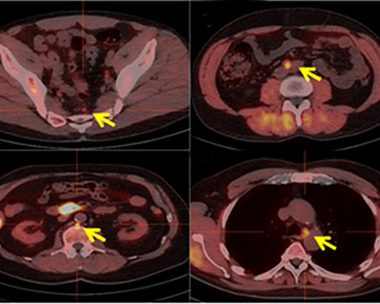
PET Scan With Axumin™ for Prostate Cancer
Lake Medical Imaging’s PET (Positron Emission Tomography) scan for prostate cancer utilizes Axumin™, an imaging agent that makes it easier to pinpoint recurring prostate cancer in patients with an elevated PSA who have been previously treated. In the past, conventional imaging tests, such as CT scans made it difficult to find prostate cancer cells that could be hidden in other parts of the body or smaller instances of the disease.
Axumin™ is a radioactive imaging agent that binds itself to prostate cancer cells in patients whose prostate cancer may have returned after treatment. The Axumin™ scan is completed within an hour with less radiation and significantly greater image resolution. With information provided by the scan, physicians can treat both the prostate and other affected areas more effectively.
Planning for your PET Scan with Axumin procedure
- Wear comfortable clothes with no metal zippers or buttons.
- Avoid exercise and do not engage in any form of strenuous or vigorous activity for 24 hours before your exam; for example, golfing, swimming, heavy lifting, and housework.
- Expect a call from our pre-registration staff who will instruct you on how to prepare for your exam.
- Plan on being at the office for approximately 1 hour (registration, injection and scan duration are included in the time approximation).
- You will be positioned on the PET scanner table and asked to lie motionless during your exam.
- During the scan, you will receive an intravenous (IV) injection of the radioactive pharmaceutical.
- The table will move slowly through the large opening of the PET scanner.
- The scan takes approximately 25 minutes.
- Usually, there are no restrictions on daily routine after the test. You should drink plenty of liquids to flush the radioactive substance from your body.
What will I experience during the procedure?
- When given the intravenous injection, you will feel a slight prick. However, you will not feel the substance in your body.
- You will be made as comfortable as possible on the exam table before you are positioned in the PET scanner for the test.
- Your technologist can see, hear and speak with you at all times during your scan.
- The small amount of radiotracer in your body will lose its radioactivity over time through the natural process of radioactive decay. Following the test, it may also pass out of your body through your urine or stool during the first few hours or days.
- You should not feel any side effects and should be able to return to your normal daily activities directly after the scan.
- We recommend that you hydrate thoroughly for 48 hours after the scan to help void the medications.
- Keep your distance from children and pregnant women for approximately six hours after a PET scan.
For more information on this topic, please visit: Radiologyinfo.org/Nuclear Medicine