
CT SCANS
The larger bore and rapid reconstruction speed of our 80- to 160-slice CT scanners provide enhanced patient comfort as well as super high-quality images. Single Energy Metal Artifact Reduction (SEMAR) technology accommodates those patients who have metal in their body, such as prosthetics. Because patient safety is always our primary concern, adaptive and integrated dose reduction strategies allow our staff to acquire images as low as reasonably achievable (ALARA), in order to minimize radiation exposure for maximum safety.
What is Computed Tomography?
A CT scan is a quick, painless procedure. Using a series of x-ray beams, a CT scanner creates cross-sectional images of the body. A computer then reconstructs these “slices” to produce a 3D image. The result is a picture with greater detail than traditional x-rays.
What are some common uses of CT?
- Studying the chest, abdomen and pelvis
- Diagnosing and treating spinal problems and injuries to the hands, feet and other skeletal structures
- Identifying injuries to the liver, spleen, kidneys, or other internal organs
- Diagnosing cancer
- Detecting, diagnosing and treating vascular diseases
- Planning and properly administering radiation treatments for tumors
- Guiding biopsies and other minimally invasive procedures
- Surgical planning
Planning for your CT procedure
- CT Abdomen and/or CT Pelvis: You must come in to pick up a prep kit at least one (1) day before your exam date
- On the day of your exam, wear comfortable, loose-fitting clothing
- Avoid clothing with zippers and snaps as metal objects can affect the image
- Depending on the part of the body that is being scanned, you may also be asked to remove hair pins, jewelry, eyeglasses, hearing aids and any dentures
- You may be asked to not eat or drink anything for one or more hours before the exam
- Inform your doctor or x-ray technologist if there is any possibility that you may be pregnant
Depending on the type of examination, contrast material may be injected through an IV, administered orally or by enema. Before administering the contrast material, you should inform the technologist of the following:
- Any allergies, especially to medications or iodine
- If you have a history of diabetes, asthma, kidney problems, heart or thyroid conditions. These conditions may indicate a higher risk of reaction to the contrast material or potential problems with eliminating the material from your system after the exam
What can I expect during this procedure?
To enhance the visibility of certain tissues or blood vessels, use of different contrast materials may be required. Depending on the type of examination, contrast material may be injected through an IV, administered orally or by enema.
The technologist positions you on the CT table. The table moves slowly into the CT scanner opening. Depending on the area of the body being examined, the increments of movement may be minimal and almost undetectable, or large enough to feel the motion. You are unaccompanied in the room during your scan, however, your technologist can see, hear and speak with you at all times.
To determine if more images are needed, you may be asked to wait until the images are reviewed.
You might feel:
- Flushed or have a metallic taste in your mouth. These are common reactions to the contrast material that disappear in a minute or two
- A warm sensation that extends to your bladder
- A mild itching sensation. If the itching persists or is accompanied by hives, it is easily treated with medication
After a CT scan with contrast, the technologist will remove the intravenous line and cover the tiny incision with a small dressing. You may resume your normal activities. A full report will be sent to your referring physician within 48 hours following your exam.
For more information on this topic, please visit: Radiologyinfo.org Computed Tomography
Using a series of X-ray beams, a CT scanner creates cross-sectional images of the body.
CT of the Head/Neck/Sinuses
CT scans of the head show bone, soft tissues, and blood vessels within the same images and can provide detailed information on head injuries and bleeding quickly enough to help save lives. This modality can also help doctors detect growths and certain diseases in the head and sinuses, evaluate “structures” such as the skull, face and sinuses for any malformations, and determine the extent of bone and soft tissue damage in patients with facial trauma to help in the planning of surgical reconstruction.
Temporal bone CT is a limited kind of CT of the head that focuses on the lower part of the skull and the surrounding soft tissues, and is often used in patients with hearing loss, chronic ear infections, and middle and inner ear diseases.
CT of the neck is done to evaluate disease in the soft tissues of the neck. Intravenous contrast is typically used during the exam to view vascular structures within the neck.
Planning for your CT of the Head/Neck/Sinuses procedure
- On the day of your exam, wear comfortable, loose-fitting clothing
- Avoid clothing with zippers and snaps as metal objects can affect the image
- Depending on the part of the body that is being scanned, you may also be asked to remove hair pins, jewelry, eyeglasses, hearing aids and any dentures
- You may be asked to not eat or drink anything for one or more hours before the exam
- Inform your doctor or x-ray technologist if there is any possibility that you may be pregnant
Contrast material may be injected through an IV, administered orally or by enema. Before administering the contrast material, you should inform the technologist of the following:
- Any allergies, especially to medications or iodine
- If you have a history of diabetes, asthma, kidney problems, heart or thyroid conditions. These conditions may indicate a higher risk of side effects to the contrast material or potential problems with eliminating the material from your system after the exam
To enhance the visibility of certain tissues or blood vessels, use of different contrast materials may be required. Depending on the type of examination, contrast material may be injected through an IV.
The technologist positions you on the CT table. The table moves slowly into the CT scanner opening. Depending on the area of the body being examined, the increments of movement may be minimal and almost undetectable, or large enough to feel the motion. You are unaccompanied in the room during your scan; however, your technologist can see, hear and speak with you at all times.
To determine if more images are needed, you may be asked to wait until the images are reviewed.
You might feel:
- Flushed or have a metallic taste in your mouth. These are common side effects to the contrast material that disappear in a minute or two
- A warm sensation that extends to your bladder
After a CT scan with contrast, the technologist will remove the intravenous line. You may resume your normal activities. A full report will be sent to your referring physician in approximately 48 hours following your exam.
CT of the Chest
CT scanning of the chest produces images that are far more detailed than a conventional chest x-ray. It is especially useful because it can simultaneously show many different types of tissue and organs including bones, soft tissues, muscle, and blood vessels of the heart and lungs.
It is helpful in the diagnosis of emphysema, COPD, pneumonia and other conditions. A contrast material is given intravenously to better visualize the vessels and organs in the chest.
Preparing for your CT of the Chest procedure
- On the day of your exam, wear comfortable, loose-fitting clothing
- Avoid clothing with zippers and snaps as metal objects can affect the image
- Depending on the part of the body that is being scanned, you may also be asked to remove hair pins, jewelry, eyeglasses, hearing aids and any dentures
- You may be asked to not eat or drink anything for one or more hours before the exam
- Inform your doctor or x-ray technologist if there is any possibility that you may be pregnant
Contrast material may be injected through an IV, administered orally or by enema. Before administering the contrast material, you should inform the technologist of the following:
- Any allergies, especially to medications or iodine
- If you have a history of diabetes, asthma, kidney problems, heart or thyroid conditions. These conditions may indicate a higher risk of side effects to the contrast material or potential problems with eliminating the material from your system after the exam
To enhance the visibility of certain tissues or blood vessels, use of different contrast materials may be required. Depending on the type of examination, contrast material may be injected through an IV.
The technologist positions you on the CT table. The table moves slowly into the CT scanner opening. Depending on the area of the body being examined, the increments of movement may be minimal and almost undetectable, or large enough to feel the motion. You are unaccompanied in the room during your scan; however, your technologist can see, hear and speak with you at all times.
To determine if more images are needed, you may be asked to wait until the images are reviewed.
You might feel:
- Flushed or have a metallic taste in your mouth. These are common side effects to the contrast material that disappear in a minute or two
- A warm sensation that extends to your bladder
After a CT scan with contrast, the technologist will remove the intravenous line. You may resume your normal activities. A full report will be sent to your referring physician in approximately 48 hours following your exam.
For more information on this topic, please visit: Radiologyinfo.org CT of the Chest
CT Lung Cancer Screening
CT lung cancer screening is a non-invasive, painless procedure that uses low-dose x-rays to screen the lungs for cancer in just 30 seconds.
A CT lung cancer screening allows the radiologist to look at different levels, or slices, of the lungs using a rotating X-ray beam. It is performed on a multi-slice spiral computed tomography (CT) scanner and can detect smaller nodules or cancer than standard chest x-rays. A tumor or nodule is a mass of extra cells that grows on the lungs. It can be benign (noncancerous) or malignant (cancerous). By detecting malignant tumors in an early stage with CT lung screening, intervention can occur at a time when the cancer is still curable and localized to the lungs.
Based on mounting evidence that lung cancer screening with CT can save lives, the American Lung Association has recommended CT lung cancer screening for smokers and former smokers.
Why is Lung Cancer Screening Important?
Lung cancer is the number one cause of cancer-related deaths in the United States. This disease is responsible for more deaths annually than breast, prostate, and colorectal cancers combined. In the United States, the lifetime risk of developing invasive lung cancer is 1 in 17 for men and 1 in 18 for women.
It is estimated that over 80% of lung cancers could be cured if detected at an early stage. Unfortunately, only 15% of lung cancers are caught at this stage, making the 5-year survival rate for all stages of lung cancer 20%. Catching lung cancer in an early stage while it is still localized to the lungs is essential. A person’s chance of survival decreases when the tumor grows to be 3 cm or more. If the cancer spreads to areas of the body outside the lungs, the survival rate is only 3%, compared with 48% if the cancer is just in the lungs. CT lung screening is extremely capable of detecting lung nodules as small as 2 or 3 cm. By catching malignant tumors when they are still small, they can be removed before disease spreads to other areas of the body.
Who Should Have Lung Cancer Screening?
Lung cancer screening is recommended for individuals between ages 50 and 80 who have any of the following risk factors:
- A family history of lung cancer
- Current smokers
- Past history of smoking (less than 10 years ago)
- Repeated exposure to secondhand smoke
- Exposure to other cancer-causing agents (e.g., asbestos and radon)
Planning for your CT of the Lung procedure
Patients lie on their backs on an exam table with arms above the head. They must hold their breath briefly as the pictures are being taken. For a short period of time, the body may be covered by a scanner, but the scanner is open at the back and front so that the patient can see out. The technologist is always able to see and hear the patient during the 15-minute procedure.
A full report will be sent to your referring physician within 48 hours following your exam.
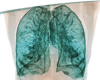

CT Lung Density “Lung Mapping” Analysis for COPD
What is CT Lung Density Analysis?
CT Lung Density Analysis (LDA) is a quantitative imaging biomarker to assist in the diagnosis and management of COPD. This groundbreaking imaging software analyzes a patient’s lung function as they inhale and exhale and enables physicians to more accurately diagnose and treat the disease. Once again, our radiologists lead the way for excellence in patient diagnoses and care.
A new non-invasive imaging technique identifies small abnormalities in the airways of patients with chronic obstructive pulmonary disease (COPD).
The technique, Parametric Response Mapping (PRM), measures patients’ lung densities as they inhale and exhale. In the past, it has traditionally been difficult to detect small airway disease early without surgery.
Planning for your CT Lung Density procedure
- Wear comfortable, loose-fitting clothing on the day of your exam.
- Avoid clothing with zippers and snaps as metal objects can affect the image.
Inform the technologist:
- If there is any possibility you are pregnant.
- Of any allergies, especially to medications or iodine.
- If you have a history of diabetes, asthma, kidney problems, heart, or thyroid conditions, since you will be injected with a contrast agent during the exam.
Plan on being with us for a minimum of 30 minutes. The technologist positions you on the CT table and pillows are used to help keep you still and in the proper position during the scan. The table moves slowly into the CT scanner opening. The increments of movement may be minimal and almost undetectable, or large enough to feel the motion.
Contrast material is injected through an IV. Before administering the contrast material, you should inform the technologist if you have:
- Any allergies, especially to medications or iodine
- A history of diabetes, asthma, kidney problems, heart or thyroid conditions. These conditions may indicate a higher risk of reaction to the contrast material or potential problems with eliminating the material from your system after the exam
You will be unaccompanied in the room during your scan; however, your technologist can see, hear, and speak with you at all times.
To determine if more images are needed, you may be asked to wait until the images are reviewed.
You might feel:
- Flushed or have a metallic taste in your mouth. These are common side effects to the contrast material that disappear in a minute or two
- A warm sensation that extends to your bladder
- If the itching persists or is accompanied by hives, it is easily treated with medication
A full report will be sent to your referring physician within 48 hours following your exam.
CT of the Abdomen/Pelvis
Using specialized equipment and expertise to create and interpret CT scans of the
abdomen and pelvis, our radiologists can accurately detect many causes of unexplained abdominal pain. CT Scans are used to visualize the liver, spleen, pancreas, kidneys, small bowel, colon and other internal organs to diagnose growths, inflammatory conditions, and in the presence of acute abdominal distress, rapidly identify emergency situations such as internal injuries and bleeding to help save lives.
Planning for your CT of the Abdomen/Pelvis procedure
- On the day of your exam, wear comfortable, loose-fitting clothing
- Avoid clothing with zippers and snaps as metal objects can affect the image
- Depending on the part of the body that is being scanned, you may also be asked to remove hair pins, jewelry, eyeglasses, hearing aids and any dentures
- You may be asked to not eat or drink anything for one or more hours before the exam
- Inform your doctor or x-ray technologist if there is any possibility that you may be pregnant
Contrast material may be injected through an IV, administered orally or by enema. Before administering the contrast material, you should inform the technologist of the following:
- Any allergies, especially to medications or iodine
- If you have a history of diabetes, asthma, kidney problems, heart or thyroid conditions. These conditions may indicate a higher risk of side effects to the contrast material or potential problems with eliminating the material from your system after the exam
The technologist positions you on the CT table. The table moves slowly into the CT scanner opening. Depending on the area of the body being examined, the increments of movement may be minimal and almost undetectable, or large enough to feel the motion. You are unaccompanied in the room during your scan; however, your technologist can see, hear and speak with you at all times.
To determine if more images are needed, you may be asked to wait until the images are reviewed.
You might feel:
- Flushed or have a metallic taste in your mouth. These are common side effects to the contrast material that disappear in a minute or two
- A warm sensation that extends to your bladder
After a CT scan with contrast, the technologist will remove the intravenous line. You may resume your normal activities. A full report will be sent to your referring physician in approximately 48 hours following your exam.
For more information on this topic, please visit: Radiologyinfo.org CT for Abdomen/Pelvis
CT of the Body/Musculoskeletal
CT is a valuable tool for taking detailed images of your musculoskeletal system, which includes your muscles, bones, joints and ligaments. It helps doctors diagnose traumatic and minor injuries as well as conditions and diseases of the bones, muscles and soft tissues. A CT scan can assist doctors to determine the exact location of damage or injury and can detect fractures that are not clear on x-rays.
Some doctors will order a CT prior to scheduled surgery to aid in their surgical planning. CT scans can also be used to monitor the effectiveness of treatments.
Planning for your CT of the Body/Musculoskeletal procedure
- On the day of your exam, wear comfortable, loose-fitting clothing
- Avoid clothing with zippers and snaps as metal objects can affect the image
- Depending on the part of the body that is being scanned, you may also be asked to remove hair pins, jewelry, eyeglasses, hearing aids and any dentures
- You may be asked to not eat or drink anything for one or more hours before the exam
- Inform your doctor or x-ray technologist if there is any possibility that you may be pregnant
Contrast material may be injected through an IV, administered orally or by enema. Before administering the contrast material, you should inform the technologist of the following:
- Any allergies, especially to medications or iodine
- If you have a history of diabetes, asthma, kidney problems, heart or thyroid conditions. These conditions may indicate a higher risk of side effects to the contrast material or potential problems with eliminating the material from your system after the exam
The technologist positions you on the CT table. The table moves slowly into the CT scanner opening. Depending on the area of the body being examined, the increments of movement may be minimal and almost undetectable, or large enough to feel the motion. You are unaccompanied in the room during your scan; however, your technologist can see, hear and speak with you at all times.
To determine if more images are needed, you may be asked to wait until the images are reviewed.
You might feel:
- Flushed or have a metallic taste in your mouth. These are common side effects to the contrast material that disappear in a minute or two
- A warm sensation that extends to your bladder
After a CT scan with contrast, the technologist will remove the intravenous line. You may resume your normal activities. A full report will be sent to your referring physician in approximately 48 hours following your exam.
CT of the Spine
The bony structure of spinal vertebrae is clearly and accurately shown by CT scanning, as are the intervertebral discs and, to some degree, the spinal cord.
CT scanning of the spine is often used to detect (or rule out) spinal damage in patients who have had injuries. CT images are ‘reconstructed’ to show many different angles of the spine and can assess congenital anomalies, such as scoliosis, or degenerative diseases.
The intervertebral discs located between the vertebrae are also evaluated and can detect disc herniations and predict whether vertebral fractures are likely to occur in patients who are at risk of osteoporosis. CT scans are helpful in evaluating the spine before and after surgery and can detect various tumors and infections in the vertebral column; and in locating areas of narrowing or compression.
Planning for your CT of the Spine procedure
- On the day of your exam, wear comfortable, loose-fitting clothing
- Avoid clothing with zippers and snaps as metal objects can affect the image
- Depending on the part of the body that is being scanned, you may also be asked to remove hair pins, jewelry, eyeglasses, hearing aids and any dentures
- You may be asked to not eat or drink anything for one or more hours before the exam
- Inform your doctor or x-ray technologist if there is any possibility that you may be pregnant
Contrast material may be injected through an IV, administered orally or by enema. Before administering the contrast material, you should inform the technologist of the following:
- Any allergies, especially to medications or iodine
- If you have a history of diabetes, asthma, kidney problems, heart or thyroid conditions. These conditions may indicate a higher risk of side effects to the contrast material or potential problems with eliminating the material from your system after the exam
The technologist positions you on the CT table. The table moves slowly into the CT scanner opening. Depending on the area of the body being examined, the increments of movement may be minimal and almost undetectable, or large enough to feel the motion. You are unaccompanied in the room during your scan; however, your technologist can see, hear and speak with you at all times.
To determine if more images are needed, you may be asked to wait until the images are reviewed.
You might feel:
- Flushed or have a metallic taste in your mouth. These are common side effects to the contrast material that disappear in a minute or two
- A warm sensation that extends to your bladder
After a CT scan with contrast, the technologist will remove the intravenous line. You may resume your normal activities. A full report will be sent to your referring physician in approximately 48 hours following your exam.
For more information on this topic, please visit: Radiologyinfo.org CT for Spine